Showing posts with label brain. Show all posts

Wednesday, October 31, 2007

Lithium and longevity?

Seems like a long shot since we still don't know how lithium works in the body, but according to Science Daily:

Nematode worms treated with lithium show a 46 percent increase in lifespan, raising the tantalizing question of whether humans taking the mood affecting drug are also taking an anti-aging medication.
Lithium is a medication that has been used to treat bipolar disorder for over 50 years. We still don't really know how it works, despite numerous studies and hypotheses.

The study above, by Buck Institute's Gordon J. Lithgow, PhD hypothesize that the process of normal aging in humans is intrinsically linked to the onset of neurodegenerative disease. They are studying hundreds of compounds for their anti-aging properties, and in the case of lithium, exploring the neuroprotective effects of lithium.

I hope they find their Fountain of Youth, but more importantly, there better be ethicists around who can wisely implement the use of it.

Friday, October 26, 2007

A brain region for addiction?

From physorg:
An animal study released Thursday bolsters the notion that drug cravings can be "switched off" by shutting down a key part of the brain, a finding that could have implications for treating addicts. Researchers have previously shown that damage to the insula can dramatically extinguish a smoker's need for nicotine.
The obvious next step is to see if this holds true for other addictions, such as as alcohol, overeating, and other drugs. Surprisingly, what researchers found in rat studies is that anesthetizing the insula also decreased unwanted medication side effects.
Chilean researchers have shown that temporarily "silencing" the insula suppresses cravings in drug-addicted rats and insulates them from the unpleasant side effects of medication, according to the study published in Science.
They hypothesize that this may be the brain region responsible for drug cravings, and that future treatments for addiction should target the insula.

Thursday, October 25, 2007

The brain area for optimism...

Is called the rostral anterior cingulate cortex (rACC).

From physorg:
The more optimistic a person is, the brighter the [rACC] area showed up in brain scans, the scientists reported in a small study published online Thursday in the journal Nature. That same part of the brain, called the rostral anterior cingulate cortex (rACC), seems to malfunction in people suffering depression, said the study co-authors, Elizabeth Phelps of New York University and Tali Sharot of University College London.
Researchers gave 15 people functional magnetic resonance imaging scans while they thought about future possibilities. When the participants thought about good events both the rACC and amygdala, which is involved in emotional responses including fear, were activated. But the correlation with optimism was biggest with the cingulate cortex.
Makes me wonder what other personality characteristics may be hard wired, or are heavily based on brain chemistry. That, then also leads to deeper philosophical questions around the nature of free will, and how much of our hopes, fears, and choices are influenced by brain chemistry and various brain regions, and how much is due to conscious choice.

Wednesday, October 24, 2007

Brain waves found that sort real memories from false

From Science Daily:
For the first time, researchers at the University of Pennsylvania are able to pinpoint brain waves that distinguish true from false memories, providing a better understanding of how memory works and creating a new strategy to help epilepsy patients retain cognitive function.
Researchers measured gamma waves and observed the following:
While patients performed the memory game, scientists observed electrical activity in their brains to determine whether specific brain waves were associated with successfully storing and retrieving memories. Researchers found that a fast brain wave, known as the gamma rhythm, increased when participants studied a word that they would later recall. The same gamma waves, whose voltage rises and fall between 50 and 100 times per second, also increased in the half-second prior to participants correctly recalling an item...
Gamma waves actually predicted whether or not an item that was about to be recalled was previously studied, said Michael Kahana, a professor of psychology
in Penns School of Arts and Sciences and lead investigator. In other words, one could see a difference in brain activity just prior to remembering something that had and had not actually happened.
I wonder how useful this could be as a lie detector?
The study will be published in November 2007's journal Psychological Science.

Tuesday, October 23, 2007

Sleep deprivation mimics psychiatric disorders

From Science Daily:

In the first neural investigation into what happens to the emotional brain without sleep, results from a brain imaging study suggest that while a good night's rest can regulate your mood and help you cope with the next day's emotional challenges, sleep deprivation does the opposite by excessively boosting the part of the brain most closely connected to depression, anxiety and other psychiatric disorders.
Anyone who's experienced a sleepless night can tell you how much harder it is for them to concentrate on basic tasks the next day, not to mention emotional regulation. Researchers at UC Berkeley's Sleep and Neuroimaging Laboratory studied the following:

Using functioning Magnetic Resonance Imaging (fMRI), Walker and his team found that the amygdala, which is also a key to processing emotions, became hyperactive in response to negative visual stimuli - mutilated bodies, children with tumors and other gory images - in study participants who stayed awake for 35 hours straight. Conversely, brain scans of those who got a full night's sleep in their own beds showed normal activity in the amygdala. "The emotional centers of the brain were over 60 percent more reactive under conditions of sleep deprivation than in subjects who had obtained a normal night of sleep," Walker said.
I wonder if someday we'll give most people a prescription for a good night's rest before starting them on antidepressants? Hopefully we'll eventually have a test we can give to patients to determine whether their psychiatric illness will get better with sleep and a vacation, or whether we need psychotropic medicatons.
Either way, here's at least another study that supports my encouragement of a good night's sleep for everyone.

Reference: Yoo et al.: "The human emotional brain without sleep -- a prefrontal amygdale disconnect." Publishing in Current Biology, Vol. 17, No. 20, R877-R878, Oct. 23, 2007

Tuesday, October 2, 2007

Mind reading computers?

It's a long way off, but the framework is beginning.

From Science Daily:
Tufts University researchers are developing techniques that could allow computers to respond to users' thoughts of frustration -- too much work -- or boredom--too little work. Applying non-invasive and easily portable imaging technology in new ways, they hope to gain real-time insight into the brain's more subtle emotional cues and help provide a more efficient way to get work done.
They are using:

functional near-infrared spectroscopy (fNIRS) technology that uses light to monitor brain blood flow as a proxy for workload stress a user may experience when performing an increasingly difficult task...

The fNIRS device, which looks like a futuristic headband, uses laser diodes to send near-infrared light through the forehead at a relatively shallow depth--only two to three centimeters--to interact with the brain's frontal lobe. Light usually passes through the body's tissues, except when it encounters oxygenated or deoxygenated hemoglobin in the blood. Light waves are absorbed by the active, blood-filled areas of the brain and any remaining light is diffusely reflected to the fNIRS detectors. "fNIRS, like MRI, uses the idea that blood flow changes to compensate for the increased metabolic demands of the area of the brain that's being used," said Erin Solovey, a graduate researcher at the School of Engineering.

So far, all they have been able to measure reliably is no workload, low workload, or high workload. However, if they are able to fine tune this tool, it could someday be used to control workflow based on the user's brain patterns. It should at least make for a nice biofeedback tool.

Tuesday, September 18, 2007

Controlling brain messengers to someday rewire the brain?

From physorg:

Researchers at MIT's Picower Institute for Learning and Memory have found that tiny, spontaneous releases of the brain's primary chemical messengers can be regulated, potentially giving scientists unprecedented control over how the brain is wired...
J. Troy Littleton, Fred and Carole Middleton Associate Professor of Biology at MIT, and colleagues found that the miniscule events that follow a burst of electrical and chemical activity among neurons are far more important that previously thought. A breakdown in this molecular mechanism could be the culprit in schizophrenia and other neurological diseases, the authors reported.
Apparently, complexins play a key part in controlling the release of the brain's chemical messengers. To study this, the team genetically modified fruit flies who make no complexins whatsoever. They determined that complexins are the gatekeepers that prevent the neurotransmitters from releasing prematurely. Complexins prevent unchecked cell growth.

This spontaneous release in the brain is not only important for signaling, it can trigger synaptic growth," Littleton said. "What's really exciting is that complexin's activity may be regulated. If we can regulate this machinery, we may be able to promote synaptic growth and potentially allow targeted rewiring in areas of the brain affected in various neurological diseases."

Wednesday, September 12, 2007

Balance enhancing ear implant

Pic: Della Santina holding multichannel vestibular prosthesis.

Scientists at the Vestibular Neuroengineering Laboratory at Johns Hopkins University are developing a vestibular prosthesis.

They have tested the concept on chincillas with some success. Usable human versions may still be years away, but the proof of concept is promising.

More from the John Hopkins University Gazette:
In its report in the June edition of the journal I.E.E.E. Transactions on Biomedical Engineering, the Johns Hopkins team showed that a matchbox-size prototype device, weighing less than three ounces, effectively mimics the workings of the inner ear's three semicircular canals by sensing head rotation and transmitting that information to the brain.
Adapting the design of cochlear implants...researchers constructed a circuit that could measure and transmit 3-D balance information to the brain through multiple electrodes connected to the vestibular nerve.
The device...consists of a head- mounted battery-operated box containing the sensors, which are positioned outside the head so that the sensors are parallel to the animal's actual semicircular canals, where head rotation is normally sensed. The sensors are connected to a microprocessor and up to eight electrodes surgically implanted in the inner ear and separately connected to nerve endings. Each electrode can act as one information channel.
Della Santina says that people disabled by loss of vestibular sensation often feel chronically off balance and lose the ability to keep the eyes steadily pointed at an object when they move their head, "seeing the world like the wobbly image on a shaky handheld video camera."
... this is the first implantable device made with multiple sensors and channels of processing that can measure and encode head rotation in all directions.
Each of the three sensors... can measure the speed of head rotation about one of three axes, or directional planes.

Tuesday, September 11, 2007

Political differences may reflect different cognitive mechanisms?

That's one hypothesis suggested by researchers at UCLA and New York University. Seems like an awfully charged statement, but a wonderful summary of the study described below.

Here's what they did (Science Daily):
NYU's David Amodio, a professor of psychology and the study's lead author, and his colleagues recorded electrical activity from the brain using electroencephalograms (EEGs) in people who rated themselves as either conservative or liberal. During these recordings, subjects had to press a button when they saw a cue, which was presented often enough that the button-press became habitual.
However, subjects occasionally saw another, infrequent cue signaling them to withhold their habitual button press. When such response inhibition was required, liberals had significantly greater neural activity originating in the anterior cingulate cortex, a portion of the brain known to be involved in conflict monitoring. Liberals were also more likely to withhold their habitual response when they saw the infrequent cue.
Previous studies have found that conservatives tend to be more persistent in their judgments and decision-making, while liberals are more likely to be open to new experiences. These differences are related to a process known as conflict monitoring-a mechanism for detecting when a habitual response is not appropriate for a new situation.

Tuesday, September 4, 2007

A treatment for dyslexia?

Using fMRI, researchers at UW's Learning Disabilities Center are studying brain connectivity timing for dyslexia.

Science Daily: "Some brain regions are too strongly connected functionally in children with dyslexia when they are deciding which sounds go with which letters," said Todd Richards, a UW neuroimaging scientist and lead author of a study published in the current issue of the Journal of Neurolinguistics. We had hints in previous studies that the ability to decode novel words improves when a specific brain region in the right hemisphere decreases in activation. This study suggests that the deactivation may result in a disconnection in time from the comparable region in the left hemisphere, which in turn leads to improved reading. Reading requires sequential as well as simultaneous processes."

So they studied a group of dyslexic kids, and a group of 'good readers'. Amazingly,

The children's brains were scanned and then those with dyslexia participated in a three-week program that taught the children the code for connecting letters and sounds with an emphasis on timing. Then the children's brains were scanned again.
Following the treatment, the fMRI scans showed that the patterns of temporal connectivity in brains of the dyslexic children had normalized and were similar to those of the good readers and spellers.


The study has shown these gains sustained for up to 2 years so far, but stresses this is not yet a cure for dyslexia. At least they're off to a good start.
How is this happening? They hypothesize:

"These results might mean that after special teaching the children with dyslexia activated letters in written words first and then switched to sounds in spoken words rather than simultaneously activating both letters and sounds," said Richards. "The overconnection between the language conductor and working memory at the same time may be a signal that working memory is overtaxed. When language processing is more efficient after treatment, working memory does not have to work as hard.

Monday, August 27, 2007

Musical perception (take a test!) and tone deafness

Apparently, there are some measurable music 'areas' in the brain.

From Science Daily:
In a study comparing amusics to people with normal musical ability, researchers used a brain imaging and statistical technique to measure the density of the white matter (which consists of connecting nerve fibers) between the right frontal lobe, where higher thinking occurs, and the right temporal lobes, where basic processing of sound occurs. The white matter of the amusics was thinner, which suggests a weaker connection. Moreover, the worse the tone deafness, the thinner the white matter.

To participate in a music perception test study online, check out http://www.delosis.com/listening developed by researchers at the University of Newcastle-upon-Tyne in England.

This site also has tests for rhythm and adaptive pitch: http://tonometric.com/adaptivepitch/

Wednesday, August 22, 2007

Magnetic brain scans to diagnose common brain diseases in 60 seconds

From Science Daily:

By comparing the patterns of tiny magnetic charges in healthy brains to those afflicted with common diseases such as Alzheimer's, the team has been able to identify the patterns commonly associated with these debilitating diseases [multiple sclerosis, Alzheimer's and schizophrenia].

How?
A process called magnetoencephalography (MEG), a non-invasive measurement of magnetic fields in the brain, has been used to examine a total of 142 volunteers during tests which last between 45-60 seconds. The team first studied 52 volunteers to find patterns of neural activity that could identify all the different illnesses. They then tested a further 46 patients to see whether the patterns found from the first group could accurately diagnose disease within a second group. Here, many of the predictors found from the first set of participants also correctly diagnosed more than 90% of subjects in the second sample.

The theory?

All behavior and cognition in the brain involves networks of nerves continuously interacting--these interactions occur on a millisecond by millisecond basis. The MEG has 248 sensors that record the interactions in the brain on a millisecond by millisecond basis, much faster than current methods of evaluation such as the functional magnetic resonance imaging (fMRI), which takes seconds to record. The measurements they recorded represent the workings of tens of thousands of brain cells.
Wow. I hope it works out, this would be an awesome tool for diagnosing, and thereby helping us choose treatments for some difficult to treat diseases.

Thursday, August 16, 2007

Depressed brains are just wired differently

From Science Daily:
In what may be the first study to use brain imaging to look at the neural circuits involved in emotional control in patients with depression, researchers at the University of Wisconsin-Madison have found that brains of people with clinical depression react very differently than those of healthy people when trying to cope with negative situations.
Apparently, non-depressed people were much better at regulating their negative emotions than depressed people. In depressed people, the harder they worked, the worse they got.
In their words:
In nondepressed individuals, high levels of regulatory activity correlated with low activity in the emotional response centers - in effect, the healthy subjects' efforts successfully quelled their emotional responses. In depressed patients, however, high levels of activity in the amygdala and other emotional centers persisted despite intense activity in the regulatory regions.
This finding suggests that healthy people are able to effectively regulate their negative emotions through conscious effort, but that the necessary neural circuits are dysfunctional in many patients with depression, the researchers say. The difference becomes even more pronounced the harder the patients try.
What does this mean? Some depressed people may not get better from cognitive therapy. If thinking about something gets someone too worked up, there will need to be other ways of helping them to get better.

I wonder how depressed people successfully managed on antidepressants would do in this study?

Friday, August 3, 2007

Neural damage by Ecstasy increased by higher ambient temperature

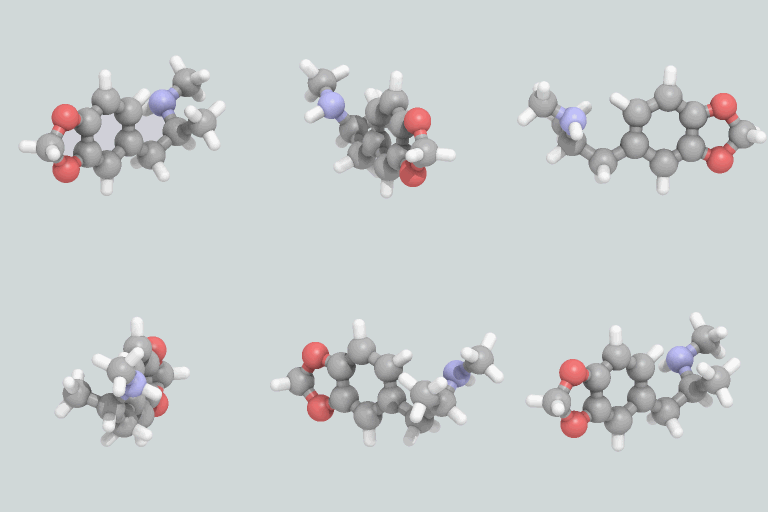
Image:MDMAanimate.gif Pic: Animated image of MDMA.

MDMA, or ecstasy is a popular 'party drug' with a fascinating history. Currently, it is most often associated with raves.

From Science Daily:
There exists a direct relationship between the consumption of MDMA, or Ecstasy, at a high ambient temperature and an increase in the neural damage which this drug provokes. This was the conclusion of the research carried out by Beatriz Goñi at the School of Pharmacy of the University of Navarra.
The study was done on rats who were given MDMA:

...at ambient temperatures of 15, 21 and 30 degrees centigrade. After performing the pertinent analyses, she demonstrated that metabolism of Ecstasy is accelerated by higher ambient temperatures at the time of administration. In addition, higher ambient temperatures also increase, in the same proportion, the neurochemical deficit that affects the brains of the users of this drug.
Unfortunately, MDMA is already documented to have several health risks. Especially of concern in in psychiatry is the long term damage to serotonin density and activity.

From wikipedia:

Serotonin is believed to play an important role in the regulation of anger, aggression, body temperature, mood, sleep, vomiting, sexuality, and appetite. Low levels of serotonin may be associated with several disorders, namely increase in aggressive and angry behaviors, clinical depression, obsessive-compulsive disorder (OCD), migraine, irritable bowel syndrome, tinnitus, fibromyalgia, bipolar disorder, anxiety disorders[citation needed] and intense religious experiences[1].
Tough to treat, too.

Thursday, August 2, 2007

Another benefit of mental and physical exercise: Decreasing schizophrenia symptoms?

As if we didn't already have enough reasons to exerise our minds and bodies.

From physorg:
Dr Anthony Hannan, along with Dr Caitlin McOmish, Emma Burrows and colleagues, characterised a genetically altered mouse and discovered that it had schizophrenia-like behaviours, including learning and memory problems, the inability to process complex information, and abnormal responses to particular sensory stimuli.
The scientists found the mouse’s condition significantly improved by simply giving them enhanced mental and physical exercise – putting running wheels in their cages, plus interesting items to smell, see and touch.
Not only did the mouse’s schizophrenia-like symptoms ease through this environmental enrichment, but a specific chemical transmitter pathway found to be abnormal in the cerebral cortex of the mice was selectively rescued.
This makes sense, since we know that schizophrenia is influenced by both genetic and environmental factors. For identical twins (twins with identical DNA), if one twin is schizophrenic, the other twin has a 50% chance of also becoming schizophrenic.

Wednesday, August 1, 2007

Using classical music and fMRI to learn how the brain pays attention

Video: This 20-second clip of a subject's fMRI illustrates how cognitive activity increases in anticipation of the transition points between movements.

Fascinating study that opens up the door to many more studies in this area.

Researchers from Stanford's School of Medicine used fMRI, and classical baroque symphonies by William Boyce (1711-79) to study event segmentation.

Event segmentation is the brain's attempt to make sense of the continual flow of information the real world generates, and how the brain partitions information into meaningful chunks by extracting information about beginnings, endings and the boundaries between events (physorg).

From Stanford's News Release:
The research team showed that music engages the areas of the brain involved with paying attention, making predictions and updating the event in memory. Peak brain activity occurred during a short period of silence between musical movements - when seemingly nothing was happening.
Here's what they observed:
An event change - the movement transition signaled by the termination of one movement, a brief pause, followed by the initiation of a new movement - activates the first network, called the ventral fronto-temporal network. Then a second network, the dorsal fronto-parietal network, turns the spotlight of attention to the change and, upon the next event beginning, updates working memory. "The study suggests one possible adaptive evolutionary purpose of music," said Jonathan Berger, PhD, professor of music and a musician who is another co-author of the study. Music engages the brain over a period of time, he said, and the process of listening to music could be a way that the brain sharpens its ability to anticipate events and sustain attention.
Makes for a great argument that people who like to sit and stare at the wall might actually be doing a whole lot of brain processing in those moments of silence and seeming inactivity.

Friday, July 27, 2007

Marijuana increases risk of psychosis

Sounds like the data finds a correlation between marijuana and psychosis, but no provable explanations for why this is the case, or whether the marijuana use actually causes the psychosis.

From physorg:
Zammit and colleagues from the University of Bristol, Imperial College and Cambridge University examined 35 studies that tracked tens of thousands of people for periods ranging from one year to 27 years to examine the effect of marijuana on mental health.
They looked for psychotic illnesses as well as cognitive disorders including delusions and hallucinations, bipolar disorder, depression, anxiety, neuroses and suicidal tendencies. They found that people who used marijuana had roughly a 40 percent higher chance of developing a psychotic disorder later in life. The overall risk remains very low.
"The strongest case is that there are consistencies across all of the studies," and that the link was seen only with psychoses - not anxiety, depression or other mental health problems, he said.

Thursday, July 26, 2007

Brain anatomy correlated to ease of learning a second language

In particular, the size of the left Heschl's Gyrus (HG), a finger-shaped structure in both the right and left side of the brain which contains the auditory cortex, was highly accurate in predicting ease of second language learning.

More details of the studies as explained from Science Daily:
The three studies have identified behavioral, neurophysiologic and, with the current study, neuroanatomic factors which, when combined, can better predict second language learning success than can each single factor alone.
In a behavioral study, Wong's group found that musical training started at an early age contributed to more successful spoken foreign language learning. The study participants with musical experience also were found to be better at identifying
pitch patterns before training.
In a neurophysiologic study -- again with the same participants -- Wong's team used functional magnetic resonance imaging to observe what parts of brain were activated when participants listened to different pitch tones. They found that the more successful second language learners were those who showed activation in the auditory cortex (where HG resides).
The participants all were native American English speakers with no knowledge of tone languages. In tone languages (spoken by half the world's population), the meaning of a word can change when delivered in a different pitch tone. In Mandarin, for example, the word "mi" in a level tone means "to squint," in a rising tone means "to bewilder" and in a falling and then rising tone means "rice."
Results:
As a group - and sometimes in fewer than two or three sessions -- the nine participants predicted on the basis of left HG size to be "more successful learners" achieved an average of 97 percent accuracy in identifying the pseudo words. The "less successful" participants averaged 63 percent accuracy and sometimes required as many as 18 training sessions to correctly identify the words.
I wonder when the HG stops growing, and/or how predictive this is in children or infants?

Tuesday, July 24, 2007

Ketamine may be the key to faster acting antidepressants?

Current antidepressants take an average of 4 to 8 weeks to relieve symptoms of depression.

Researchers have been experimenting with ketamine with interesting results.

Ketamine [from wikipedia] is a dissociative anesthetic for use in human and veterinary medicine. Ketamine has a wide range of effects in humans, including analgesia, anesthesia, hallucinations, arterial hypertension, and bronchodilation.[3] It is primarily used for the induction and maintenance of general anesthesia, usually in combination with some sedative drug. Other uses include sedation in intensive care, analgesia (particularly in emergency medicine), and treatment of bronchospasm. It is also a popular anesthetic in veterinary medicine.

Because of these side effects, it has also become a somewhat popular recreational drug.

Here's the latest from physorg:
A new study has revealed more about how the medication ketamine, when used experimentally for depression, relieves symptoms of the disorder in hours instead of the weeks or months it takes for current antidepressants to work...
Ketamine blocks a receptor called NMDA on brain cells, an earlier NIMH study in humans had shown, but the new study in mice shows that this is an intermediate step. It turns out that blocking NMDA increases the activity of another receptor, AMPA, and that this boost in AMPA is crucial for ketamine’s rapid antidepressant actions. The study was reported online in Biological Psychiatry on July 23, by NIMH researchers Husseini K. Manji, MD, Guang Chen, MD, PhD, Carlos Zarate, MD, and colleagues.
Sounds like it may still be several years down the line, but being able to have antidepressants that take hours instead of months to work would make my job much easier. Seems that we may also have a new mechanism of antidepressant action that focuses on AMPA and the glutamate system(an inhibitory neurotransmitter) instead of serotonin.

Writer cramp due to brain abnormalities?

What exactly is writer's cramp? From wikipedia:
Writer's cramp is a form of cramp or spasm that affects certain muscles of the hand and fingers as a result of excessive fine motor activity like writing or playing the piano. It is referred to medically as task-specific focal dystonia of the hand.
From physorg:

People with serious cases of writer’s cramp have brain abnormalities, according to a study published in the July 24, 2007, issue of Neurology, the medical journal of the American Academy of Neurology. People with writer’s cramp had less brain tissue than healthy people in three areas of the brain that connect the senses and movement with their affected hand...

The researchers found that those with writer’s cramp had less grey matter in three areas of the brain: the cerebellum, the thalamus, and the sensorimotor cortex.

At least there are computers, so those with writer's cramp can see if they are any more or less susceptible to carpal tunnel.